
What hurts?
Some possible conditions • Foot Pain – Inside of Foot – Rearfoot
-
 Sprain/Strain
 -
 Adult Acquired Flatfoot
Some possible conditions • Foot Pain – Inside of Foot – Rearfoot
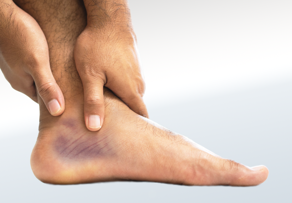
Ankle sprains and strains have similar symptoms, causes and risk factors, but they also have some differences. Ankle sprains are usually a result of a traumatic or acute injury, while ankle strains can be acute or chronic. Acute injuries of the ankle occur suddenly and are caused by a single episode, whereas a chronic injury tends to develop after a period of time with repetitive movement or overuse. The ankle joint is formed by the tibia in the lower leg and the talus bone in the foot. There are dozens of ligaments to support the ankle and several muscles and tendons to provide strength and stability. If any of the ligaments are overstretched or disrupted, an ankle sprain may result. When the muscles or tendons are affected, a strain can occur. In either case, the discomfort is typically localized to the ankle and rearfoot. An eversion ankle injury typically causes issues on the inside of the rearfoot/ankle.
The main difference between a sprain and strain is the onset of the pain, swelling, redness and reduced range of motion at the ankle. When an ankle sprain occurs, there may be an audible pop or a popping feeling, followed by bruising at the site and intense pain at the time of the injury. A strain will typically occur over time and can also result in weakness and instability around the ankle joint. A strain can also occur from a one-time injury or possibly at the same time as a sprain. Sprains and strains are graded from 1 to 3 and based on severity. Severity ranges from minor tearing to complete tearing of the soft tissues. Symptoms will be worse the higher the grade.
Ankle sprains and strains have similar risk factors including overtraining causing ligament or muscle fatigue, poor athletic conditioning, being overweight, not warming up sufficiently before training, poor and unsupportive footwear and history of a previous ankle injury. Another ankle injury is much more likely to occur after it has already been injured due to a sprain or strain. Additionally, women over the age of 30 and young men between the ages of 15 and 24 tend to be more at risk for ankle sprains. Activities and sports carried out on uneven terrain or involving pivoting on the spot or side-to-side movement may also increase a person’s risk for injury at the ankle.
Healing after an ankle sprain can be lengthy as ligaments have little to no blood supply and knowing if ankle pain is a result of a sprain or strain is imperative for proper treatment and avoiding re-injury. Mild ankle sprains and strains can be treated at home with R.I.C.E, the age old application of rest, ice, compression and elevation. Ankle joint instability is common after injury. Ankle braces and taping can be effective for increasing stability, as are custom foot orthotics and appropriate footwear. Stretching and strengthening exercises will help to support and stabilise the weakened ankle joint. More severe injuries may also require immobilisation of the joint with traditional plaster casts or an air cast/boot. Surgery to repair the injured structures is only required if the first-line of treatment and rehabilitation is ineffective.
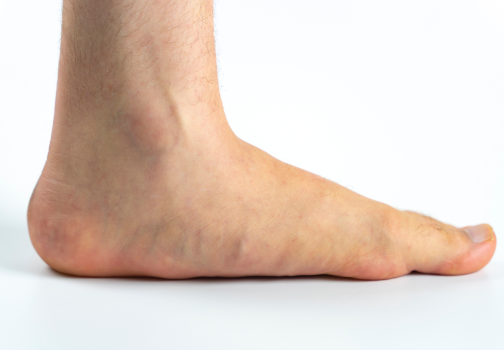
Adult acquired flatfoot, also referred to as posterior tibial tendon dysfunction (PTTD), is characterized by a fallen arch and outward pointed toes. This condition is a gradual progression in most cases, as the posterior tibialis (PT) tendon becomes weakened over time and loses the ability to maintain the height of the arch. Adult acquired flatfoot is different from a flatfoot condition in children. Adults with flatfeet remain permanently flat without surgical intervention, while children can outgrow the condition on their own, sometimes with or without treatment.
Those with adult acquired flatfoot deformity (AAFD) typically present with pain along the course of the posterior tibialis tendon, which is on the inside of the foot and ankle. Pain is usually worse with activities, especially high impact, like running and jumping. The heel bone can shift toward the outside of the foot putting more pressure on the outside ankle bone. Patients that develop arthritis may also have nerve impingement (trapping/pinching of the nerve), which can lead to numbness, tingling or burning in the arch and toes of the foot. Diabetic patients may notice swelling or a bump on the bottom of the foot if the condition progresses. Due to the presence of diabetic neuropathy (loss of feeling in their feet) in some patients, pain is not always an indicator of this condition. But, for most people, tenderness, achiness, soreness of the tendon and inner ankle are hallmark signs, alongside a seemingly low or flattened arch on the inside of the foot.
The most common cause of AAFD/PTTD is damage to the posterior tibial tendon. This tendon starts at the calf and travels down to the inside of the foot. If the tendon becomes swollen, irritated or torn, the arch will begin to collapse due to lack of support and strength from the tendon. Studies have shown that women over 40 years of age are the most likely to develop an issue with the PT tendon. Risk factors that can increase the chances of AAFD/PTTD include but are not limited to obesity, diabetes, and high blood pressure. There is a small population of patients with inflammatory arthritis, such as rheumatoid arthritis, that develop a painful flatfoot condition. Inflammatory arthritis typically causes dysfunction of the tendon, ligaments and joints leading to a more severe flatfoot deformity. Diabetic patients can progress to a condition called Charcot foot, which entails severe flattening and a “rocker bottom” like deformity on the bottom of the midfoot. Bones can break and coalesce as the arch collapses, requiring an even more specialized brace in order to maintain weight bearing.
Patients with adult acquired flatfoot deformity are often treated with orthotics, footwear and braces. They are cared for by a Canadian Certified Pedorthist or Registered Chiropodist. A biomechanical examination is recommended in these cases due to the complex nature of the deformity. Often patients require nonsteroidal anti-inflammatory medication due to pain and swelling, which can limit mobility. Patients also benefit from physical therapy to help treat and prevent further injuries with the tendon involved. Those that do not respond to conservative therapy may require surgical intervention.


