
What hurts?
Some possible conditions • Ankle Pain – Ankle – Back
-
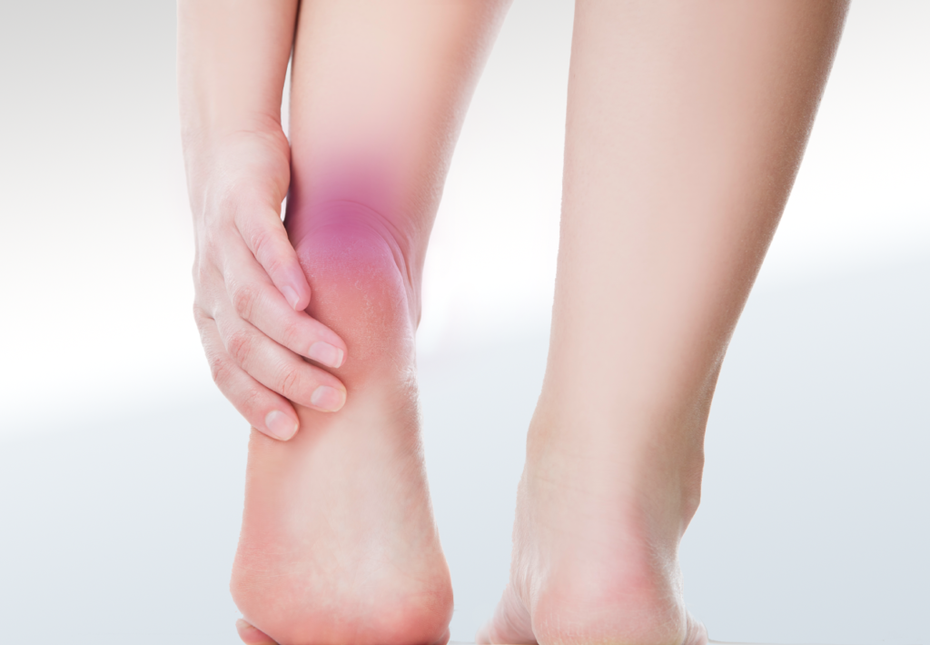 Sever’s Apophysitis
 -
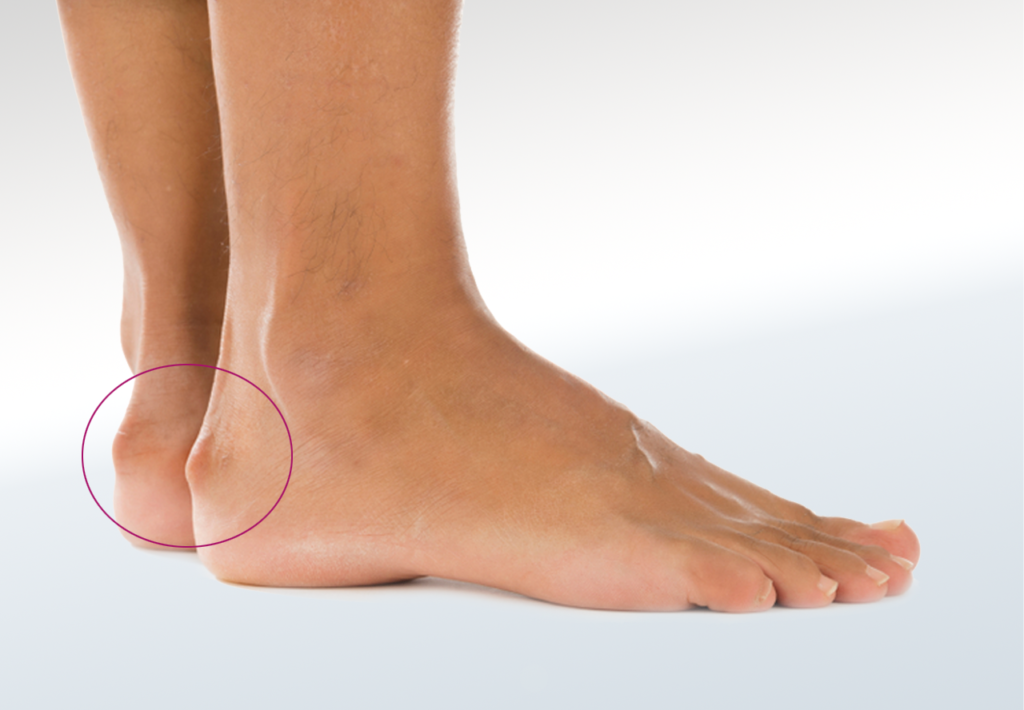 Haglunds
 -
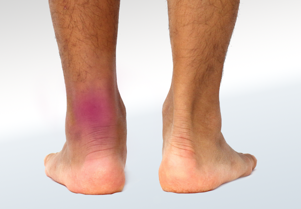 Achilles Rupture/Tear
 -
 Achilles Tendinitis/Tendinosis
Some possible conditions • Ankle Pain – Ankle – Back
In growing children, the most common cause of heel pain is Sever’s Disease, or calcaneal apophysitis. This condition begins when the growth plate that is at the back of the heel, or calcaneus, becomes inflamed and painful. Children that are active and play sports or exercise regularly have a higher likelihood of developing the condition.
Children often present with pain and swelling on the back of the heel. There can be increased warmth to the area as well. Pain touching the area is often indicative of Sever’s disease. Pain to the sides and bottom of the heel are also not uncommon. Limping after running or jumping and stiffness after sitting for long periods or sleeping are typical presentations too.
The cause of Sever’s disease is repetitive stress on the growth plate at the back of the heel bone, where the Achilles tendon attaches and pulls. Since patients undergoing a rapid growth spur are more susceptible to developing inflammation at the growth plate, the typical ages of presentation in girls is 8 to 13 and boys 10 to 15. The growth plate is a soft area of cartilage where bone growth occurs. The Achilles tendon connects the calf muscles to the heel bone in the area of the growth plate. Activities that increase the pull of the Achilles tendon on the heel, such as running and jumping, can irritate the heel growth plate. Overuse and improper shoes are also causes for possible growth plate injuries.
First and foremost in order to treat Sever’s disease, rest and time off of intense activities is a must. Gentle stretching of the calf muscles can help reduce the stress on the heel, but may not be tolerated when acutely painful. Physical therapy plays a large role in the stretching and strengthening of the leg muscles and tendons. An evaluation with a Chiropodist or Pedorthist is recommended due to the development of the child. Icing the heel may be recommended to decrease the present inflammation. A Chiropodist can evaluate the use of over-the-counter nonsteroidal anti-inflammatory medications to help reduce pain and swelling of the growth plate. A Canadian Certified Pedorthist evaluation is helpful in order to adjust shoes or offer advice on footwear and provide orthotics with heel lifts that will cushion the heel and reduce tension to the Achilles tendon.
Haglund’s deformity, also known as retrocalcaneal exostosis or a “pump bump”, is a very common condition of the heel. The deformity is caused by an abnormality of the heel bone, or calcaneus, and soft tissues of the foot. The calcaneus tends to enlarge on the back of the bone causing irritation to the soft tissues near, such as the Achilles tendon. Shoes often contribute to the pain and irritation if rub begins due to the enlargement of the bone on the back of the heel. The use of high heel shoes coined the term “pump bump” due to the irritation caused from tight stiff dress shoes and rubbing while walking.
The most common age for presentation is 40-50 years old, and within females more than males. The symptoms are most commonly bilateral with pain at the back of the heel, more pronounced after rest. Limping and swelling are often seen with time. History of conditions such as gout, rheumatoid arthritis or seronegative spondyloarthropathies should be ruled out as they can mimic the deformity seen. You may have blistering or bursa inflammation on the back of the heel due to the enlarged bump on the back of the heel and shoes rubbing.
This condition is caused by genetics, lifestyle and shoes. But, there are factors that contribute to symptoms that present such as tight or poorly fitting shoes, altered biomechanics, and overuse, such as in runners and athletes. A tight Achilles tendon, high arch of the foot, tendency to walk on the outside of the foot, and heredity are also suggested causes of the pain and deformity.
Haglund’s deformities are more often treated conservatively than surgically. X-rays may be taken in order to fully evaluate the heel bone for enlargement Spurs where the Achilles tendon attaches to the heel bone can also be seen on X-rays. Changing the heel height of a shoe, adding orthotics or heel lifts to current footwear, anti-inflammatory medications and physical therapy are most commonly utilized. An evaluation with a Chiropodist or Pedorthist can be performed in order to evaluate shoes, gait and Achilles tendon and ankle flexibility. With acute severe pain, immobilization may be necessary with a pneumatic walker or cast, in a non-weight bearing position.
The Achilles tendon, the largest tendon in the body, connects the calf muscle to your heel. The tendon can withstand significant stresses from running and jumping, but is vulnerable to injury. Achilles tendon ruptures are defined as a separation of the tendon off of the heel bone, or transverse tear of the tendon. When a rupture of the tendon occurs, the tendon is unable to perform normal function.
Patients often describe a rupture of the Achilles tendon sounding like a “pop”, followed by an immediate sharp pain in the back of the ankle or lower leg. The ability to walk is immediately affected with the rupture. Swelling begins and is located mainly near the heel. The ability to “push off” of the injured leg or stand on tip toes is grossly affected by the injury. Your doctor or footcare clinician may note a “gap” in the tendon if it completely ruptured. A test involving compression of the calf muscle, in order to elicit plantar flexion of the foot, may also be performed in order to rule in or out a tendon rupture.
Achilles tendon ruptures are the result of a sudden injury to the tendon. The Achilles tendon aids in plantarflexion of the foot, or pointing downward, as well as the push off strength when walking, jumping or running. The area most commonly ruptured is roughly 2.5 inches from the attachment onto the heel bone. This section is known as the watershed region as it tends to have poor blood flow and impaired healing potential. Typical types of injuries include falling from a height, stepping into a hole or sudden burst of activity or intensity. Age can play a role in rupture with 30-50 year olds being the most common population. Men are more likely to have a rupture than women. Steroid injections in the ankle joint area have been shown to cause weakening of the surrounding tissues, such as tendons. There is a link between certain oral antibiotics and tendon ruptures. Unfortunately since the Achilles is the largest tendon in the body it is at an increased risk with these medications.
The treatment options for an Achilles tendon tear are dependent on the age and activity level of the patient. Younger and more active patients, such as athletes, may opt for surgical intervention, while older, less active patients may opt for nonsurgical treatment. Nonsurgical treatment has shown to have optimal results. The nonsurgical approach involves resting the tendon by staying off of the injured limb, applying ice to the area, compression to aid with swelling, and immobilization via a pneumatic boot. Patients are often placed in slight plantar flexion to reduce any strain on the tendon. Your family doctor also plays a role in treatment with prescription anti-inflammatory medications. A professional will be able to monitor healing in order to obtain full rehabilitation. There is a slight increase in chances of re-rupture with a conservative approach, however there are less risks than when surgery is involved. Lastly, physical therapy has been to be successful in rebuilding the tendon and strengthening the tendon following rupture. Following the tendon healing, it is advisable to be properly fitted for footwear and if required, orthotics, to optimize your biomechanics.
Achilles tendonitis is a common condition affecting one of the largest tendons in the body. The tendon connects the calf muscles to the heel bone and is vital in the function of walking, running, climbing stairs, jumping, and standing on tiptoes. Tendonitis occurs when the tendon becomes acutely inflamed and irritated. Tendinopathy is often associated with this condition, described as microscopic degeneration due to chronic damage over a period of time. There are two distinct types of Achilles tendonitis, insertional and non-insertional tendonitis. Unfortunately, the two types of Achilles tendonitis can occur separately or succinctly. Non-insertional Achilles tendonitis is characterized by inflammation of the fibers in the middle portion of the tendon, above the attachment to the heel bone. Whereas, insertional Achilles tendonitis involves the lower portion of the tendon as it attaches to the heel bone. The tendon fibers may calcify over time and bone spurs can form on the back of the heel.
The symptoms associated with Achilles tendonitis include pain and stiffness along the Achilles tendon particularly first thing in the morning, pain along the back of the heel that worsens with activity, and thickening of the tendon. Bone spur formation, chronic swelling and pain with shoe wear are also signs and symptoms of tendonitis. Non-insertional Achilles tendonitis is more often found in younger, and active patients. When there is palpation pain in the middle of the tendon for non-insertional or at the back of the heel bone for insertional, it’s associated with limited range-of-motion in your ankle.
Achilles tendonitis, unlike Achilles ruptures, is not usually related to a specific injury. Repetitive stress to the tendon, such as over exertion is the most common cause. Other factors that contribute to the development of Achilles tendonitis include tight calf muscles, haglund’s deformity, or a sudden increase in the intensity or amount of exercise.
Treatment for Achilles tendonitis is aimed at providing pain relief and reducing inflammation. Anti-inflammatory medication, such as ibuprofen, or prescription medication may be used to reduce the inflammation within the tendon. Initial treatment may include rest, icing, footwear modification, orthotics, stretching/physical therapy and oral medications. Physical therapy is aimed at stretching and strengthening the calf muscles and reducing stress on the Achilles tendon. A Pedorthist can fit and dispense a night splint, which holds the foot in place while in a calf stretch position while you sleep. Maintaining healthy calf flexibility will not only help treat Achilles tendonitis but will aid in preventing recurrent issues. Supportive footwear with an open back or soft heel may help to reduce the shearing and irritation of the tendon during healing. A pneumatic walking boot may be necessary for severe pain and inflammation, often a period of non-weight bearing is necessary to reduce the strain to the tendon. More advanced therapies such as extracorporeal shockwave therapy have shown to promote healing of the damaged tendon.


